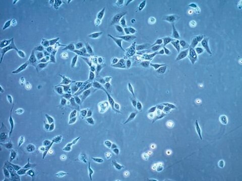
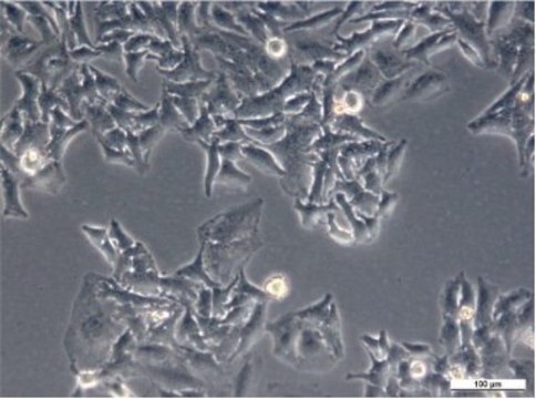
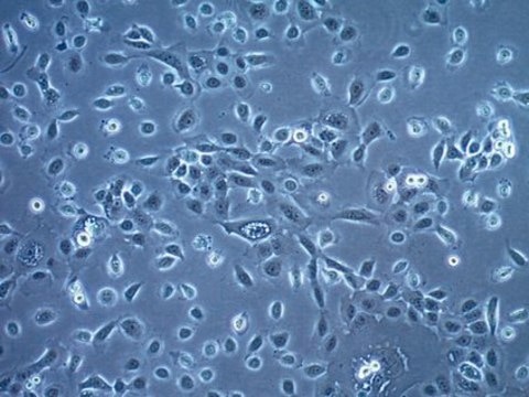
A431 Cell Line human
85090402, squamous carcinoma
Synonym(s):
A-431 Cells, A431/P Cells
About This Item
Recommended Products
biological source
human skin
growth mode
Adherent
karyotype
Hypertriploid
morphology
Epithelial
products
Not specified
receptors
Epidermal growth factor (EGF)
technique(s)
cell culture | mammalian: suitable
relevant disease(s)
cancer
shipped in
dry ice
storage temp.
−196°C
Related Categories
Cell Line Origin
Cell Line Description
Application
- to demonstrate that p38 mitogen-activated protein kinase enhances epidermal growth factor-induced apoptosis in A431 cells
- to study melanoma-specific CD44 (a tumor marker and transmembrane receptor) alternative splicing pattern in A431 cells
- to study the anti-cancer activity of antioxidant-rich Cameroonian medicinal plants
Culture Medium
Subculture Routine
Other Notes
related product
Certificates of Analysis (COA)
Search for Certificates of Analysis (COA) by entering the products Lot/Batch Number. Lot and Batch Numbers can be found on a product’s label following the words ‘Lot’ or ‘Batch’.
Already Own This Product?
Find documentation for the products that you have recently purchased in the Document Library.
Articles
Application note on how the CellASIC® ONIX2 microfluidic system can be used to analyze caspase-3 mediated apoptosis/cell death and cellular hypoxia in live immune and cancer cell lines.
Our team of scientists has experience in all areas of research including Life Science, Material Science, Chemical Synthesis, Chromatography, Analytical and many others.
Contact Technical Service


