SCC628
MNK-3 Mouse Innate Lymphoid Cell Line

Synonym(s):
MNK-3 Mouse Innate Lymphoid Cell Line, ILC3 cell line, MNK cell line, MNK3
Sign Into View Organizational & Contract Pricing
All Photos(1)
About This Item
UNSPSC Code:
41106514
Recommended Products
biological source
mouse
Quality Level
packaging
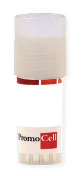
vial of ≥1 x 10^6 vial (viable cells per vial)
manufacturer/tradename
Millipore
technique(s)
cell culture | mammalian: suitable
shipped in
liquid nitrogen
storage temp.
−196°C
Application
- Each vial contains > 1X10⁶ viable cells.
- Cells are tested negative for infectious diseases by a Mouse Essential CLEAR Panel by Charles River Animal Diagnostic Services.
- Cells are verified to be of mouse origin and negative for interspecies contamination from human, rat, Chinese hamster, Golden Syrian hamster, and nonhuman primate (NHP) as assessed by a Contamination Clear panel by Charles River Animal Diagnostic Services
- Cells are negative for mycoplasma contamination.
Features and Benefits
MNK-3 is a novel ILC3 cell line which expresses the key transcription factors, cytokine secretion, cytokine receptors, chemokine receptors, and adhesion molecules represented in innate lymphoid cells.
Target description
Innate Lymphoid Cells (ILCs) contribute to the innate immune system through cell signaling and regulation of immune cells. ILCs lack functionally rearranged T-Cell Receptors (TCRs) or immunoglobulin genes.1 ILCs have been categorized into three subsets. Group 1 ILCs include Natural Killer cells (NK) and other cells that produce IFN-γ but are distinct from NK cells.1 Group 2 ILCs are classified by their capability to produce cytokines, especially IL-5 and IL-13. Group 2 ILCs are sometimes known by variable names such as innate helper type-2 cells or natural helper cells.1 Group 3 ILCs are classified by the expression of Rorγt which is also needed for their development. This classification includes Lymphoid Tissue inducer cells (LTi) in addition to adult Rorγt+ ILC cells. ILC3 cells are important for lymphoid tissue development, mucosal homeostasis, containment of commensal bacteria, inhibition of the pathology that causes inflammatory bowel disease, thymic regeneration, control of mucosal IgA production, and immune defense capabilities against bacterial and fungal infections. ILC3 cells are also an important source of IL-22 which aids in the protection of intestinal mucosa from pathogens.1 MNK-3 is a novel, prototypical ILC3 cell line model which expresses the key transcription factors, cytokine secretion, cytokine receptors, chemokine receptors, and adhesion molecules represented in ILC3 cells. The MNK parental cell line was generated from sorted NIH-Swiss mouse fetal day-15 NK1.1+ thymocytes which were later transfected with constructs encoding the SV40 large T antigen and human c-MYC. Parental MNK cells were sorted to be NK-receptor negative. Two sublines were developed from parental MNK and later designated MNK-1 and MNK-3. NKR- MNK parental cells were cultured in IL-7 and IL-15 and confirmed to be negative for CD94, NKG2D, DX5, and CD11b. These NKR- cells became the MNK-3 subline.1 MNK-3 cells display LTi-like activity in vitro when incubated with mesenchymal stromal cells. A low ICAM-1, high VCAM-1 OP9 variant cell line was co-cultured with MNK-3 cells which resulted in a significant increase in ICAM-1 expression. This is essentially used as a readout for LTi activity.1 MNK-3 cells were confirmed by ELISA to secrete IL-22 at a basal level in addition to increased IL-22 secretion when stimulated by IL-1β, IL-23, and IL-2. MNK-3 cells also serve as a model for a high number of applications presented by ILC3 functions such as IL-17A induction. MNK-3 cells are also easy to genetically modify which can aid in application function. Source Cell line generated from sorted NIH-Swiss mouse NK1.1+ thymocytes. Cells were infected with a Bcl-2 retroviral producer cell line and transfected with replication-deficient SV40 and human c-myc constructs by electroporation.References1. Allan DS, Kirkham CL, Aguilar OA, Qu LC, Chen P, Fine JH, Serra P, Awong G, Gommerman JL, Zúñiga-Pflücker JC, et al. 2014. An in vitro model of innate lymphoid cell function and differentiation. Mucosal Immunology. 8(2):340–351. doi:https://doi.org/10.1038/mi.2014.71.
Storage and Stability
MNK-3 cells should be stored in liquid nitrogen. The cells can be cultured for at least 10 passages without significantly affecting cell marker expression and function.
Other Notes
This product is intended for sale and sold solely to academic institutions for internal academic research use per the terms of the “Academic Use Agreement” as detailed in the product documentation. For information regarding any other use, please contact licensing@milliporesigma.com.
Disclaimer
Unless otherwise stated in our catalog or other company documentation accompanying the product(s), our products are intended for research use only and are not to be used for any other purpose, which includes but is not limited to, unauthorized commercial uses, in vitro diagnostic uses, ex vivo or in vivo therapeutic uses or any type of consumption or application to humans or animals.
wgk_germany
WGK 2
flash_point_f
Not applicable
flash_point_c
Not applicable
Certificates of Analysis (COA)
Search for Certificates of Analysis (COA) by entering the products Lot/Batch Number. Lot and Batch Numbers can be found on a product’s label following the words ‘Lot’ or ‘Batch’.
Already Own This Product?
Find documentation for the products that you have recently purchased in the Document Library.
Our team of scientists has experience in all areas of research including Life Science, Material Science, Chemical Synthesis, Chromatography, Analytical and many others.
Contact Technical Service


