General description
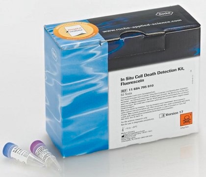
Non-isotopic kit for the detection of apoptosis at the individual cell level. Apoptotic morphology is easily detected. This kit allows the recognition of apoptotic nuclei in paraffin-embedded tissue sections, tissue cryosections, or in cell preparations fixed on slide by Fragment End Labeling (FragEL) of DNA. Terminal deoxynucleotidyl transferase (TdT) binds to exposed ends of DNA fragments generated in response to apoptotic signals and catalyzes the template-dependent addition of biotin-labeled and unlabeled deoxynucleotides. Biotinylated nucleotides are detected using streptavidin-horseradish peroxidase (HRP) conjugate. Diaminobenzidine reacts with the labeled sample to generate an insoluble colored product at the site of DNA fragmentation. Counterstaining with methyl green aids in the morphological evaluation and characterization of normal and apoptotic cells.
Components
Counterstain, Proteinase K, Equilibration and Labeling Buffers, TdT Enzyme (Cat. No. PF060), Stop Buffer, Blocking Buffer, 50X Conjugate, DAB, Positive and Negative Control Slides, H₂O₂/Urea Tablets, Methyl Green Counterstain, and a user protocol.
Warning
Toxicity: Multiple Toxicity Values, refer to MSDS (O)
Principle
The Calbiochem TdT-FragEL DNA Fragmentation Detection Kit is a non-isotopic system for the labeling of DNA breaks in apoptotic nuclei in paraffin-embedded tissue sections, tissue cryosections, and in cell preparations fixed on slides.
Preparation Note
1. Generation of Negative Control SamplesA negative control of your specific sample can be generated by substituting dH2O for the TdT in the reaction mixture or by keeping the specimen in 1X reaction buffer (with a cover slip) during the labeling step. Perform all other steps as described. This controls for endogenous peroxidases and non-specific conjugate binding. A non-apoptotic control is also critical since cells and tissue begin to undergo apoptosis from the very beginning of the excision, fixation, and processing steps. A delay in fixation or routine mechanical manipulation may result in the unwanted breakage of DNA that could be misinterpreted as apoptosis.2. Generation of Positive Control Samples: A positive control of your specific tissue sample can be generated by covering the entire specimen with 1 µg/µl DNase I in 1X TBS/1 mM MgSO4 following proteinase K treatment. Incubate at room temperature for 20 min. Perform all other steps as described. This will fragment DNA in normal cells. Cytoplasmic as well as nuclear DAB staining of DNase treated cells may be observed.
Storage and Stability
Upon arrival store the entire contents of the kit at -20°C in a non-frostfree freezer. Following initial thaw store the Stop Buffer and Methyl Green Counterstain at room temperature and the Blocking Buffer at 4°C. Refreezing of these components, however, does not appear to affect their performance.
Other Notes
Due to the nature of the Hazardous Materials in this shipment, additional shipping charges may be applied to your order. Certain sizes may be exempt from the additional hazardous materials shipping charges. Please contact your local sales office for more information regarding these charges.
Xiao, D. and Bullock, R. 1996. J. Neurosurgery85, 655.
Gu, Y., et al. 1994. Endocrinology135, 1272.
Gavrieli, Y., et al. 1992. J. Cell Biol.119, 493.
Fawthrop, D. J., et al. 1991. Arch. Toxicol.65, 437.
Martin, S.J., et al. 1990. J. Immunology145, 1859.
Wyllie, A. H. 1980. Nature284, 555.
Kerr, J. F. R., et al. 1972. Br. J. Cancer26, 239.
Legal Information
CALBIOCHEM is a registered trademark of Merck KGaA, Darmstadt, Germany

