Skip to Content
MilliporeSigma
All Photos(2)

Documents

MABS64

Sigma-Aldrich

Anti-phospho-p38 (Thr180/Tyr182) Antibody, clone 6E5.2

clone 6E5.2, from mouse

Synonym(s):

mitogen-activated protein kinase 14, cytokine suppressive anti-inflammatory drug binding protein, stress-activated protein kinase 2A, Cytokine suppressive anti-inflammatory drug-binding protein, CSAID-binding protein, MAP kinase MXI2, MAX-interacting pro

Sign Into View Organizational & Contract Pricing


About This Item

UNSPSC Code:
12352203
eCl@ss:
32160702
NACRES:
NA.41

biological source

mouse

Quality Level

antibody form

purified immunoglobulin

antibody product type

primary antibodies

clone

6E5.2, monoclonal

species reactivity

human, mouse

technique(s)

immunocytochemistry: suitable
western blot: suitable

NCBI accession no.

UniProt accession no.

shipped in

wet ice

target post-translational modification

phosphorylation (pThr180/pTyr182)

Gene Information

human ... MAPK14(1432)

General description

The p38 family refers to the SAPK2/3 Kinases (Stess-Activated Protein Kinases), a subfamily of JNK/SAPK family. p38/SAPK Kinases are also activated by stresses, most notably inflammatory cytokines, irradiation, and certain toxins such as anisomycin and arsenite. SAPKs have been implicated in many pathological conditions, including inflammation, cancer and neurodegenerative diseases. As with the other MAPK family members (ERK, JNK, p38MAPK, and ERK5), p38 is activated through phosphorylation on both threonine and tyrosine residues at the Thr-X-Tyr dual phosphorylation motif, where X is Gly for p38. Once phosphorylated and active, it then goes on to phosphorylate downstream targets in both the cytoplasm and the nucleus. Some of their main targets of p38 are the kinase MAPKAPK2 and the transcription factor ATF2 that are activated via their phosphorylation.

Specificity

This antibody recognizes p38 when phosphorylated at Thr180 and Tyr182.

Immunogen

Epitope: Phosphorylated Thr180 and Tyr182
Linear peptide corresponding to human p38 phosphorylated at Thr180 and Tyr182.

Application

Detect phospho-p38 (Thr180/Tyr182) using this Anti-phospho-p38 (Thr180/Tyr182) Antibody, clone 6E5.2 validated for use in WB & IC.
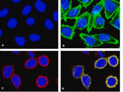
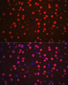
Immunocytochemistry Analysis: 1:500 dilution from a previous lot detected p38 in Anisomycin treated NIH/3T3 cells.
Research Category
Signaling
Research Sub Category
MAP Kinases

Quality

Evaluated by Western Blot in Anisomycin treated and untreated HeLa cell lysate.

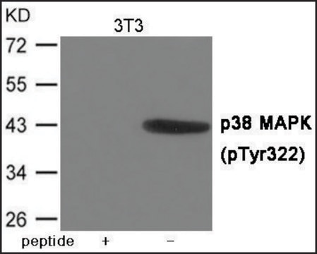
Western Blot Analysis: 0.5 µg/mL of this antibody detected p38 on 10 µg of Anisomycin treated and untreated HeLa cell lysate.

Target description

41 kDa observed

Physical form

Format: Purified
Protein G Purified
Purified mouse monoclonal IgGκ in buffer containing 0.1 M Tris-Glycine (pH 7.4, 150 mM NaCl) with 0.05% sodium azide.

Storage and Stability

Stable for 1 year at 2-8°C from date of receipt.

Analysis Note

Control
Anisomycin treated and untreated HeLa cell lysate

Other Notes

Concentration: Please refer to the Certificate of Analysis for the lot-specific concentration.

Disclaimer

Unless otherwise stated in our catalog or other company documentation accompanying the product(s), our products are intended for research use only and are not to be used for any other purpose, which includes but is not limited to, unauthorized commercial uses, in vitro diagnostic uses, ex vivo or in vivo therapeutic uses or any type of consumption or application to humans or animals.

Not finding the right product?  

Try our Product Selector Tool.

Storage Class

12 - Non Combustible Liquids

wgk_germany

WGK 1

flash_point_f

Not applicable

flash_point_c

Not applicable


Certificates of Analysis (COA)

Search for Certificates of Analysis (COA) by entering the products Lot/Batch Number. Lot and Batch Numbers can be found on a product’s label following the words ‘Lot’ or ‘Batch’.

Already Own This Product?

Find documentation for the products that you have recently purchased in the Document Library.

Visit the Document Library

Simona Daniele et al.
Cellular signalling, 27(8), 1609-1629 (2015-04-26)
Growing evidence suggests that alterations of the inflammatory/immune system contribute to the pathogenesis of major depression and that inflammatory processes may influence the antidepressant treatment response. Depressed patients exhibit increased levels of inflammatory markers in both the periphery and brain
Jili Wen et al.
Molecular medicine reports, 16(5), 7548-7552 (2017-09-26)
Infections by pathogens may lead to cardiovascular diseases, including acute/chronic myocarditis. (Coxsackieviruses B3) CVB3 is considered to be the most common causative agent in m‑yocarditis, which can lead to dilated cardiomyopathy. The present study aimed to investigate the mechanism of
Weiyong Shen et al.
Glia, 62(7), 1110-1124 (2014-04-02)
Retinal diseases such as macular telangiectasis type 2 (MacTel), age-related macular degeneration (AMD) and diabetic retinopathy (DR) affect both neurons and blood vessels. Treatments addressing both at the same time might have advantages over more specific approaches, such as vascular

Our team of scientists has experience in all areas of research including Life Science, Material Science, Chemical Synthesis, Chromatography, Analytical and many others.

Contact Technical Service