Skip to Content
MilliporeSigma
All Photos(1)

Documents

MABC1158

Sigma-Aldrich

Anti-phospho-MLKL (Ser345) Antibody, clone 7C6.1

clone 7C6.1, from mouse

Synonym(s):

Mixed lineage kinase domain-like protein

Sign Into View Organizational & Contract Pricing


About This Item

UNSPSC Code:
12352203
eCl@ss:
32160702
NACRES:
NA.43

biological source

mouse

Quality Level

antibody form

purified antibody

antibody product type

primary antibodies

clone

7C6.1, monoclonal

species reactivity

mouse

technique(s)

ELISA: suitable
immunocytochemistry: suitable
western blot: suitable

isotype

IgG2bκ

NCBI accession no.

UniProt accession no.

shipped in

ambient

target post-translational modification

phosphorylation (pSer345)

Gene Information

mouse ... Mlkl(74568)

General description

Mixed lineage kinase domain-like protein (UniProt: Q9D2Y4; also known as MLKL) is encoded by the Mlkl gene (Gene ID: 74568) in murine species. MLKL is a pseudokinase that plays a key role in TNF-induced necroptosis. MLKL is highly expressed in thymus, colon, intestine, liver, spleen and lung and is expressed at much lower level in skeletal muscle, heart and kidney. It is not detected in brain. MLKL is activated following phosphorylation by RIPK3 that induces a conformational switch, which is essential for necroptosis. The protein kinase domain of MLKL is catalytically inactive, but contains a pseudoactive site with an interaction between Lys-219 and Gln-343 residues. Upon phosphorylation by RIPK3, MLKL undergoes an active conformation and homotrimerization through its amino-terminal coiled-coil domain. Phosphorylation is also essential for homotrimerization of MLKL and its subsequent localization to the plasma membrane. Plasma membrane localization of MLKL is considered to be essential for Ca2+ influx, which is an early event of TNF-induced necroptosis. (Ref.: Cai, Z et al. (2014). Nat. Cell Biol. 16(1): 55-65).

Specificity

Clone 7C6.1 specifically detects MLKL phosphorylalated on Ser345. It targets a sequence of 19 amino acids surrounding Ser345 in the C-terminal region.

Immunogen

KLH-conjugated linear peptide corresponding to 19 amino acids surrounding Ser345 from the C-terminal half of murine Mixed lineage kinase domain-like protein (MLKL).

Application

Detect phospho MLKL Ser345 using this mouse monoclonal Anti-phospho-MLKL (Ser345), clone 7C6.1, Cat. No. MABC1158. It has been tested in ELISA, Immunocytochemistry, and Western Blotting.
ELISA Analysis: A representative lot detected phospho-MLKL (Ser345) in ELISA applications (Rodriguez, D.A., et. al. (2016). 23(1):76-88).

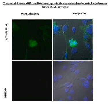
Immunocytochemistry Analysis: A representative lot detected phospho-MLKL (Ser345) in Immunocytochemistry applications (Rodriguez, D.A., et. al. (2016). 23(1):76-88).

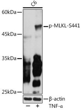
Western Blotting Analysis: A representative lot detected phospho-MLKL (Ser345) in Western Blotting applications (Rodriguez, D.A., et. al. (2016). 23(1):76-88).
Research Category
Apoptosis & Cancer

Quality

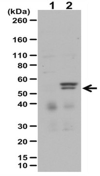
Evaluated by Western Blotting in MLKL -/- mouse embryonic fibroblasts expressing doxycycline-inducible wild-type MLKL and treated with TNF alpha and Z-VAD-FMK.

Western Blotting Analysis: A 1:4,000 dilution of this antibody detected phospho-MLKL Ser345 in 20 ug of lysate from MLKL -/- mouse embryonic fibroblasts expressing doxycycline-inducible wild-type MLKL-Flag-C construct and treated with TNF alpha (10 ng/mL) and Z-VAD-FMK (25 uM).


Target description

~54 kDa observed; 54.32 kDa calculated. Uncharacterized bands may be observed in some lysate(s).

Physical form

Format: Purified
Protein G purified
Purified mouse monoclonal antibody IgG2b in buffer containing 0.1 M Tris-Glycine (pH 7.4), 150 mM NaCl with 0.05% sodium azide.

Storage and Stability

Stable for 1 year at 2-8°C from date of receipt.

Other Notes

Concentration: Please refer to lot specific datasheet.

Disclaimer

Unless otherwise stated in our catalog or other company documentation accompanying the product(s), our products are intended for research use only and are not to be used for any other purpose, which includes but is not limited to, unauthorized commercial uses, in vitro diagnostic uses, ex vivo or in vivo therapeutic uses or any type of consumption or application to humans or animals.

Not finding the right product?  

Try our Product Selector Tool.

wgk_germany

WGK 1

flash_point_f

Not applicable

flash_point_c

Not applicable


Certificates of Analysis (COA)

Search for Certificates of Analysis (COA) by entering the products Lot/Batch Number. Lot and Batch Numbers can be found on a product’s label following the words ‘Lot’ or ‘Batch’.

Already Own This Product?

Find documentation for the products that you have recently purchased in the Document Library.

Visit the Document Library

Carlos J Nogueras-Ortiz et al.
Cells, 9(7) (2020-07-09)
We have previously shown that blood astrocytic-origin extracellular vesicles (AEVs) from Alzheimer's disease (AD) patients contain high complement levels. To test the hypothesis that circulating EVs from AD patients can induce complement-mediated neurotoxicity involving Membrane Attack Complex (MAC) formation, we
Sulayman Benmerzoug et al.
Nature communications, 9(1), 5226-5226 (2018-12-13)
Silica particles induce lung inflammation and fibrosis. Here we show that stimulator of interferon genes (STING) is essential for silica-induced lung inflammation. In mice, silica induces lung cell death and self-dsDNA release in the bronchoalveolar space that activates STING pathway.
François Sipieter et al.
iScience, 24(9), 103074-103074 (2021-09-28)
ERK1/2 involvement in cell death remains unclear, although many studies have demonstrated the importance of ERK1/2 dynamics in determining cellular responses. To untangle how ERK1/2 contributes to two cell death programs, we investigated ERK1/2 signaling dynamics during hFasL-induced apoptosis and
Xuan Li et al.
Cell death discovery, 7(1), 338-338 (2021-11-10)
Necroptosis, a form of programmed cell death, accounts for many inflammations in a wide range of diseases. Diet-induced obesity is manifested by low-grade inflammation in the mediobasal hypothalamus (MBH), and microglia are implicated as critical responsive components for this process.

Our team of scientists has experience in all areas of research including Life Science, Material Science, Chemical Synthesis, Chromatography, Analytical and many others.

Contact Technical Service