Skip to Content
MilliporeSigma
All Photos(2)

Documents

ABT45

Sigma-Aldrich

Anti-Claudin-5 Antibody

from rabbit, purified by affinity chromatography

Synonym(s):

Claudin-5, Transmembrane protein deleted in VCFS, TMDVCF

Sign Into View Organizational & Contract Pricing


About This Item

UNSPSC Code:
12352203
eCl@ss:
32160702
NACRES:
NA.41

biological source

rabbit

Quality Level

antibody form

affinity isolated antibody

antibody product type

primary antibodies

clone

polyclonal

purified by

affinity chromatography

species reactivity

rat, mouse, horse, human

species reactivity (predicted by homology)

equine (based on 100% sequence homology), ox (based on 100% sequence homology)

packaging

antibody small pack of 25 μg

technique(s)

immunohistochemistry: suitable (paraffin)
western blot: suitable

NCBI accession no.

UniProt accession no.

shipped in

ambient

storage temp.

2-8°C

target post-translational modification

unmodified

Gene Information

human ... CLDN5(7122)

Related Categories

General description

Claudin-5, a transmembrane protein deleted in velo-cardio-facial syndrome (TMVCF), seems to be involved in endothelial tight junctions. Claudin-5 has been exclusively found in the cell-cell borders of endothelial cells. Lower claudin-5 levels in sinusoidal endothelial cells could lead to their dysfunction during liver injuries. Levels of claudin-5 in tumor vessels could be a potential marker for hepatocellular carcinoma prognosis. The loss of claudin-5 function has been linked to carcinogenesis.

Specificity

This antibody recognizes Claudin-5 at the cytoplasmic domain.

Immunogen

Epitope: Cytoplasmic Domain
KLH-conjugated linear peptide corresponding to human Claudin-5 at the cytoplasmic domain.

Application

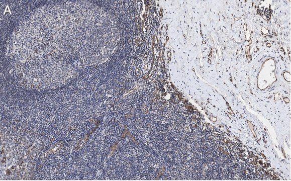
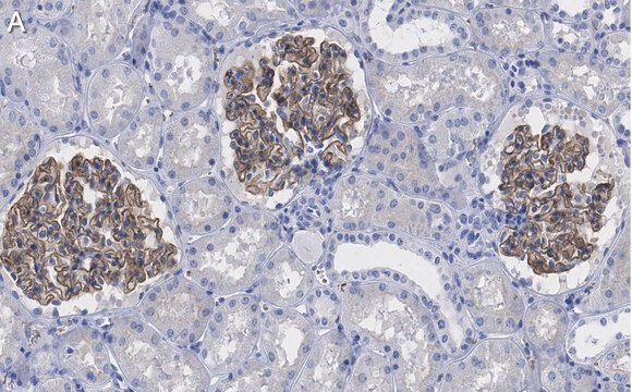
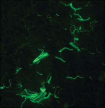
Anti-Claudin-5 Antibody detects level of Claudin-5 & has been published & validated for use in WB, IH(P).
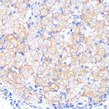
Immunohistochemistry Analysis: A 1:800 dilution from a representative lot detected Clausin-5 in prostate tissue.
Research Category
Cell Structure
Research Sub Category
Adhesion (CAMs)

Quality

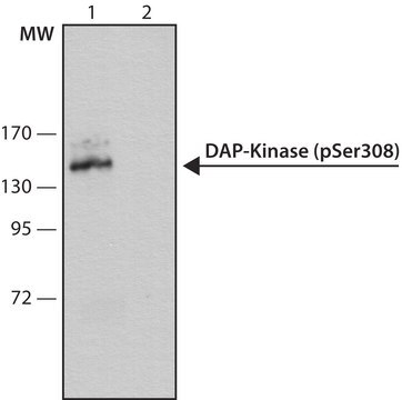
Evaluated by Western Blot in human lung tissue lysate.

Western Blot Analysis: 0.1 µg/mL of this antibody detected Claudin-5 in 10 µg of human lung tissue lysate.

Target description

~17 kDa observed.
Molecular weights at ~17 kDa and 62 kDa may be observed depending on phosphorylation and glycosylation effects (Ishizaki, T., et al. (2003). Experimental Cell Research. 290:275–288).

Physical form

Affinity purified
Purified rabbit polyclonal in buffer containing 0.1 M Tris-Glycine (pH 7.4), 150 mM NaCl with 0.05% sodium azide.

Storage and Stability

Stable for 1 year at 2-8°C from date of receipt.

Analysis Note

Control
Human lung tissue lysate

Other Notes

Concentration: Please refer to the Certificate of Analysis for the lot-specific concentration.

Disclaimer

Unless otherwise stated in our catalog or other company documentation accompanying the product(s), our products are intended for research use only and are not to be used for any other purpose, which includes but is not limited to, unauthorized commercial uses, in vitro diagnostic uses, ex vivo or in vivo therapeutic uses or any type of consumption or application to humans or animals.

Not finding the right product?  

Try our Product Selector Tool.

Storage Class

12 - Non Combustible Liquids

wgk_germany

WGK 1

flash_point_f

Not applicable

flash_point_c

Not applicable


Certificates of Analysis (COA)

Search for Certificates of Analysis (COA) by entering the products Lot/Batch Number. Lot and Batch Numbers can be found on a product’s label following the words ‘Lot’ or ‘Batch’.

Already Own This Product?

Find documentation for the products that you have recently purchased in the Document Library.

Visit the Document Library

Haitao Liu et al.
Diabetologia, 62(12), 2365-2374 (2019-10-16)
Levels of neutrophil elastase, a serine protease secreted by neutrophils, are elevated in diabetes. The purpose of this study was to determine whether neutrophil elastase (NE) contributes to the diabetes-induced increase in retinal vascular permeability in mice with streptozotocin-induced diabetes
Disrupted tight junctions in the small intestine of cystic fibrosis mice.
De Lisle, RC
Cell and Tissue Research null
Zhi Fang et al.
FASEB journal : official publication of the Federation of American Societies for Experimental Biology, 30(6), 2097-2107 (2016-02-19)
The mechanism of blood-brain barrier (BBB) disruption, involved in poststroke edema and hemorrhagic transformation, is important but elusive. We investigated microRNA-150 (miR-150)-mediated mechanism in the disruption of BBB after stroke in rats. We found that up-regulation of miR-150 increased permeability
Hongyan Zhu et al.
Neurochemical research, 43(10), 1897-1904 (2018-08-09)
Blood-brain barrier (BBB) destruction is associated with a variety of neurological diseases. Brain microvascular endothelial cells (BMECs) are the key constituent of BBB. Both matrix metalloproteinases-2/9 (MMP-2/9) and toll-like receptor-2 (TLR2) are coexpressed in BMECs and have been shown to
Karsten Bartels et al.
Heart, lung & circulation, 23(10), 981-984 (2014-06-17)
Neurologic injury is common after cardiac surgery and disruption of the blood brain barrier (BBB) has been proposed as a contributing factor. We sought to study BBB characteristics in a rodent model of cardiopulmonary bypass (CPB) and deep hypothermic circulatory

Our team of scientists has experience in all areas of research including Life Science, Material Science, Chemical Synthesis, Chromatography, Analytical and many others.

Contact Technical Service