Skip to Content
MilliporeSigma
All Photos(1)

Documents

AB2988

Sigma-Aldrich

Anti-LYVE1 Antibody

from rabbit, purified by affinity chromatography

Synonym(s):

Cell surface retention sequence-binding protein 1, Extracellular link domain-containing protein 1, Hyaluronic acid receptor, cell surface retention sequence binding protein-1, extracellular link domain containing 1, extracellular link domain-containing 1

Sign Into View Organizational & Contract Pricing


About This Item

UNSPSC Code:
12352203
eCl@ss:
32160702
NACRES:
NA.41

biological source

rabbit

Quality Level

antibody form

affinity isolated antibody

antibody product type

primary antibodies

clone

polyclonal

purified by

affinity chromatography

species reactivity

human

species reactivity (predicted by homology)

mouse (based on 100% sequence homology)

technique(s)

immunohistochemistry: suitable

isotype

IgG

NCBI accession no.

UniProt accession no.

shipped in

wet ice

target post-translational modification

unmodified

Gene Information

human ... LYVE1(10894)

General description

LYVE1 has been identified as a major receptor for HA (extracellular matrix glycosaminoglycan hyaluronan) on the lymph vessel wall. The deduced amino acid sequence of LYVE1 predicts a 322-residue type I integral membrane polypeptide 41% similar to the CD44 HA receptor with a 212-residue extracellular domain containing a single Link module the prototypic HA binding domain of the Link protein superfamily. Like CD44, the LYVE1 molecule binds both soluble and immobilized HA. However, unlike CD44, the LYVE1 molecule colocalizes with HA on the luminal face of the lymph vessel wall and is completely absent from blood vessels. Hence, LYVE1 is the first lymph-specific HA receptor to be characterized and is a uniquely powerful marker for lymph vessels themselves. LYVE1 may function in lymphatic hyaluronan transport. Additionally, LYVE1 has a role in tumor metastasis.

Specificity

Rat (90% sequence homology).
This antibody recognizes LYVE1.

Immunogen

Epitope: Unknown
KLH-conjugated linear peptide corresponding to mouse LYVE1.

Application

Anti-LYVE1 Antibody is an antibody against LYVE1 for use in IH.
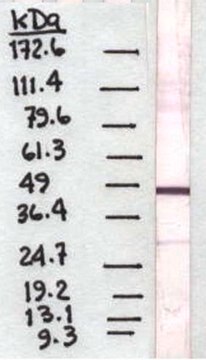
Not recommended to work in western blot using mouse liver lysate.
Research Category
Apoptosis & Cancer
Research Sub Category
Tumor Markers

Quality

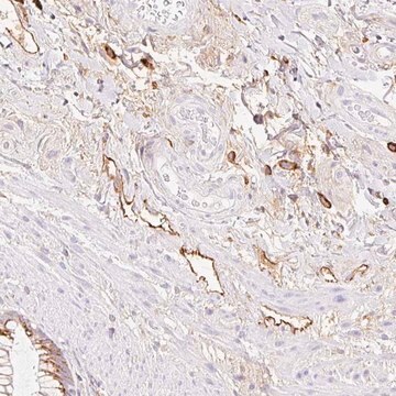
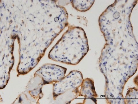
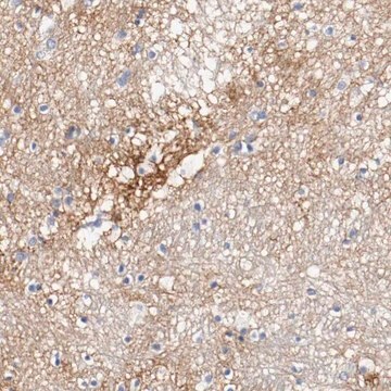
Evaluated by Immunohistochemistry in ovarian cancer tissue.

Immunohistochemistry Analysis: 1:1,000 dilution of this antibody detected LYVE1 in ovarian cancer tissue.

Target description

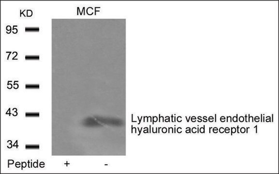
~ 40 kDa

Linkage

Replaces: 07-538

Physical form

Affinity purified
Purified rabbit polyclonal in buffer containing 0.1 M Tris-Glycine (pH 7.4, 150 mM NaCl) with 0.05% sodium azide.

Storage and Stability

Stable for 1 year at 2-8°C from date of receipt.

Analysis Note

Control
Ovarian cancer tissue

Other Notes

Concentration: Please refer to the Certificate of Analysis for the lot-specific concentration.

Disclaimer

Unless otherwise stated in our catalog or other company documentation accompanying the product(s), our products are intended for research use only and are not to be used for any other purpose, which includes but is not limited to, unauthorized commercial uses, in vitro diagnostic uses, ex vivo or in vivo therapeutic uses or any type of consumption or application to humans or animals.

Still not finding the right product?  

Give our Product Selector Tool a try.

Storage Class

12 - Non Combustible Liquids

wgk_germany

WGK 1

flash_point_f

Not applicable

flash_point_c

Not applicable


Certificates of Analysis (COA)

Search for Certificates of Analysis (COA) by entering the products Lot/Batch Number. Lot and Batch Numbers can be found on a product’s label following the words ‘Lot’ or ‘Batch’.

Already Own This Product?

Find documentation for the products that you have recently purchased in the Document Library.

Visit the Document Library

Ssu-Han Wang et al.
International journal of cancer, 144(11), 2795-2810 (2018-11-30)
Oral squamous cell carcinoma (OSCC) LN1-1 cells previously showed greater capacities for lymphangiogenesis and lymph node metastasis compared to their parental OEC-M1 cells, in addition to an ability to enhance the migration and tube formation of lymphatic endothelial cells (LECs).
Abdi Ghaffari et al.
Breast cancer research : BCR, 16(5), 438-438 (2014-09-19)
Recent evidence suggests that tumour lymphangiogenesis promotes lymph node metastasis, a major prognostic factor for survival of breast cancer patients. However, signaling mechanisms involved in tumour-induced lymphangiogenesis remain poorly understood. The expression of ezrin, a membrane cytoskeletal crosslinker and Src

Our team of scientists has experience in all areas of research including Life Science, Material Science, Chemical Synthesis, Chromatography, Analytical and many others.

Contact Technical Service