Skip to Content
Merck
All Photos(4)

Documents

P8825

Sigma-Aldrich

Monoclonal Anti-Proliferating Cell Nuclear Antigen antibody produced in mouse

clone PC 10, ascites fluid

Synonym(s):

Monoclonal Anti-Proliferating Cell Nuclear Antigen, Anti-PCNA

Sign Into View Organizational & Contract Pricing


About This Item

UNSPSC Code:
12352203
NACRES:
NA.41

biological source

mouse

Quality Level

conjugate

unconjugated

antibody form

ascites fluid

antibody product type

primary antibodies

clone

PC 10, monoclonal

contains

15 mM sodium azide

species reactivity

human, monkey, insect, yeast
rat, mouse

packaging

antibody small pack of 25 μL

technique(s)

flow cytometry: suitable
immunocytochemistry: suitable
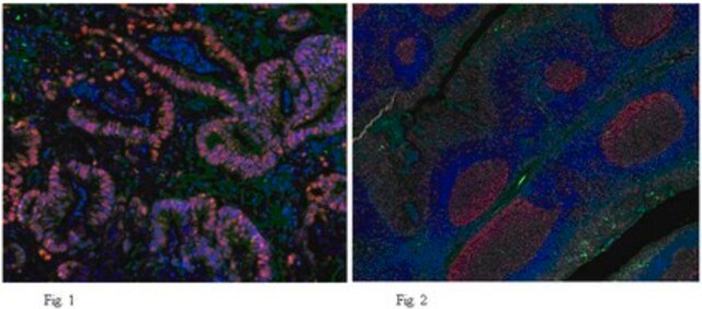
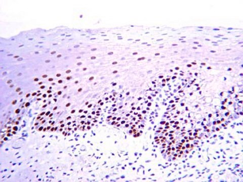
immunohistochemistry (formalin-fixed, paraffin-embedded sections): 1:3,000 using human tonsil sections
immunohistochemistry (frozen sections): suitable
immunoprecipitation (IP): suitable
indirect ELISA: suitable
microarray: suitable
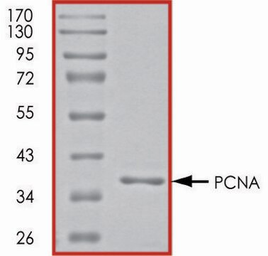
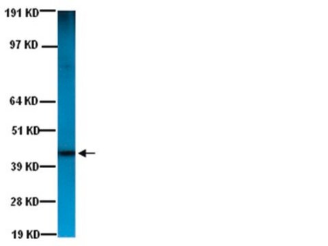
western blot: 1:3,000 using HS-68 human foreskin- cell extract

isotype

IgG2a

UniProt accession no.

shipped in

dry ice

storage temp.

−20°C

target post-translational modification

unmodified

Gene Information

human ... PCNA(5111)

Looking for similar products? Visit Product Comparison Guide

General description

Monoclonal Anti-Proliferating Cell Nuclear Antigen (mouse IgG2a isotype) is derived from the PC 10 hybridoma produced by the fusion of mouse myeloma cells and splenocytes from a BALB/c mouse immunized with PCNA-Protein A fusion protein. Proliferating cell nuclear antigen (PCNA, 36 kDa), also known as cyclin, is an auxiliary protein of DNA polymerase. The protein is present in the nucleoplasm of continually cycling cells throughout the cell cycle.
PCNA (proliferating cell nuclear antigen) is a member of the structurally and functionally conserved family of DNA sliding clamps (β clamps). They exist as ring-shaped complexes- homotrimers in eukaryotes, having pseudohexameric symmetry. A monomer of PCNA is composed of two similar globular domains, connected by an interdomain connecting loop, which is a long, and probably a flexible loop. These monomers organize themselves in a head-to-tail manner to form a ring, which has an inner positively charged surface composed of α helices and an outer surface of β sheets.

Specificity

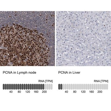
Recognizes the acidic, non-histone, auxiliary protein of DNA polymerase, PCNA, also known as polymerase delta accessory protein. Fixation duration can markedly affect the intensity of PCNA immunoreactivity. However, delay in fixation does not affect the immunoreactivity. Enzymatic treatment destroys staining. In immunocytochemical labeling of acetone-methanol, or methanol-fixed cells, the antibody shows granular staining throughout the nucleus (nucleolus and nucleoplasm). Specific staining is observed in proliferating cell nuclei, particularly in germinal centers, of a wide range of normal and malignant tissues.

Immunogen

PCNA-Protein A fusion protein

Application

Monoclonal Anti-Proliferating Cell Nuclear Antigen antibody produced in mouse has been used for immunohistochemistry (IHC) Western blotting (WB).
Monoclonal Anti-Proliferating Cell Nuclear Antigen antibody produced in mouse has been used in:
  • enzyme linked immuno sorbent assay (ELISA)
  • immunoblotting
  • immunohistology
  • immunoprecipitation
  • flow cytometry

Biochem/physiol Actions

PCNA (proliferating cell nuclear antigen) is loaded around the template-primer 3′ ends, which is recognized by the conserved chaperone-like complex RFC (replication factor C), and this mechanism is ATP-dependent. Encircling of the DNA by the PCNA ring ensures firm anchoring of the polymerases to the DNA, and hence, functions as a co-factor for DNA polymerases. Post-translation modifications of this protein at the K164 residue is required for the regulation of DNA damage tolerance (DDT) pathways, pathways that ensure recovery from replication arrest at DNA lesions.
Proliferating cell nuclear antigen (PCNA) is essential for DNA replication during S-phase. It is essential for cellular DNA synthesis. PCNA is required for leading strand synthesis in the SV40 system where it acts as an auxiliary protein for polymerase.

Disclaimer

Unless otherwise stated in our catalog or other company documentation accompanying the product(s), our products are intended for research use only and are not to be used for any other purpose, which includes but is not limited to, unauthorized commercial uses, in vitro diagnostic uses, ex vivo or in vivo therapeutic uses or any type of consumption or application to humans or animals.

Not finding the right product?  

Try our Product Selector Tool.

Storage Class Code

12 - Non Combustible Liquids

WGK

nwg

Flash Point(F)

Not applicable

Flash Point(C)

Not applicable


Certificates of Analysis (COA)

Search for Certificates of Analysis (COA) by entering the products Lot/Batch Number. Lot and Batch Numbers can be found on a product’s label following the words ‘Lot’ or ‘Batch’.

Already Own This Product?

Find documentation for the products that you have recently purchased in the Document Library.

Visit the Document Library

Polycystic kidney disease in Han:SPRD Cy rats is associated with elevated expression and mislocalization of SamCystin.
Nagao S et al
American Journal of Physiology: Renal Physiology, 299(5), F1078-F1086 (2010)
PCNA, the maestro of the replication fork.
Moldovan GL et al
Cell, 129(4), 665-679 (2007)
Proliferating cell nuclear antigen (PCNA): ringmaster of the genome
Paunesku T, et al.
International Journal of Radiation Biology, 77(10), 1007-1021 (2001)
Relevance of simultaneous mono-ubiquitinations of multiple units of PCNA homo-trimers in DNA damage tolerance.
Kanao R et al
PLoS ONE, 10(2), e0118775-e0118775 (2015)
Development of cerebellar neurons and glias revealed by in utero electroporation: Golgi-like labeling of cerebellar neurons and glias.
Kita Y et al
PLoS ONE, 8(7), e70091-e70091 (2013)

Our team of scientists has experience in all areas of research including Life Science, Material Science, Chemical Synthesis, Chromatography, Analytical and many others.

Contact Technical Service