Skip to Content
Merck
All Photos(4)

Documents

HPA011866

Sigma-Aldrich

Anti-PCDH7 antibody produced in rabbit

Prestige Antibodies® Powered by Atlas Antibodies, affinity isolated antibody, buffered aqueous glycerol solution

Synonym(s):

Anti-BH-Pcdh, Anti-PPP1R120

Sign Into View Organizational & Contract Pricing


About This Item

UNSPSC Code:
12352203
Human Protein Atlas Number:
NACRES:
NA.41

biological source

rabbit

Quality Level

conjugate

unconjugated

antibody form

affinity isolated antibody

antibody product type

primary antibodies

clone

polyclonal

product line

Prestige Antibodies® Powered by Atlas Antibodies

form

buffered aqueous glycerol solution

species reactivity

human

technique(s)

immunohistochemistry: 1:200- 1:500

immunogen sequence

SGEVTFSLESGSEYLKIDNLTGELSTSERRIDREKLPQCQMIFDENECFLDFEVSVIGPSQSWVDLFEGQVIVLDINDNTPTFPSPVLTLTVEENRPVGTLYLLPT

UniProt accession no.

shipped in

wet ice

storage temp.

−20°C

target post-translational modification

unmodified

Gene Information

human ... PCDH7(5099)

Immunogen

Protocadherin-7 precursor recombinant protein epitope signature tag (PrEST)

Application

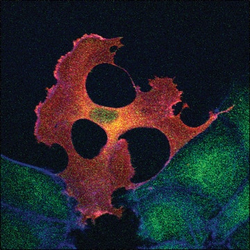
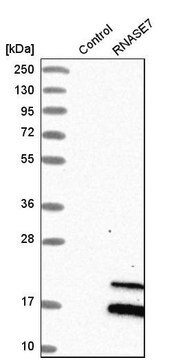
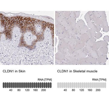
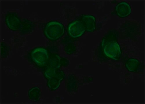
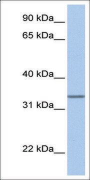
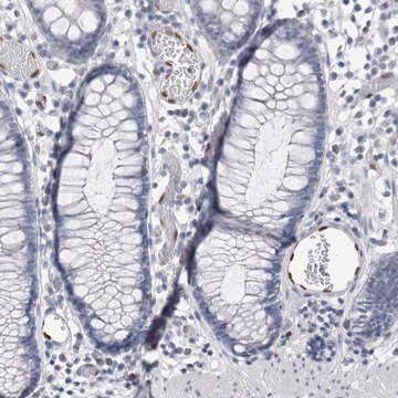
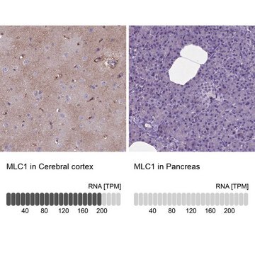
Anti-PCDH7 antibody produced in rabbit, a Prestige Antibody, is developed and validated by the Human Protein Atlas (HPA) project . Each antibody is tested by immunohistochemistry against hundreds of normal and disease tissues. These images can be viewed on the Human Protein Atlas (HPA) site by clicking on the Image Gallery link. The antibodies are also tested using immunofluorescence and western blotting. To view these protocols and other useful information about Prestige Antibodies and the HPA, visit sigma.com/prestige.

Biochem/physiol Actions

PCDH7 (Protocadherin 7) encodes a protocadherin belonging to the cadherin superfamily. It is predominantly expressed in cerebral cortex neurons of the brain. Pcdh7 performs an essential role in osteoclastogenesis by promoting cell-cell fusion. Upregulated PCDH7 expression has been documented in studies, which indicate an important role of PCDH7 in bone metastasis of breast cancer.

Features and Benefits

Prestige Antibodies® are highly characterized and extensively validated antibodies with the added benefit of all available characterization data for each target being accessible via the Human Protein Atlas portal linked just below the product name at the top of this page. The uniqueness and low cross-reactivity of the Prestige Antibodies® to other proteins are due to a thorough selection of antigen regions, affinity purification, and stringent selection. Prestige antigen controls are available for every corresponding Prestige Antibody and can be found in the linkage section.

Every Prestige Antibody is tested in the following ways:
  • IHC tissue array of 44 normal human tissues and 20 of the most common cancer type tissues.
  • Protein array of 364 human recombinant protein fragments.

Linkage

Corresponding Antigen APREST71519

Physical form

Solution in phosphate-buffered saline, pH 7.2, containing 40% glycerol and 0.02% sodium azide

Legal Information

Prestige Antibodies is a registered trademark of Merck KGaA, Darmstadt, Germany

Disclaimer

Unless otherwise stated in our catalog or other company documentation accompanying the product(s), our products are intended for research use only and are not to be used for any other purpose, which includes but is not limited to, unauthorized commercial uses, in vitro diagnostic uses, ex vivo or in vivo therapeutic uses or any type of consumption or application to humans or animals.

Not finding the right product?  

Try our Product Selector Tool.

WGK

WGK 1

Flash Point(F)

Not applicable

Flash Point(C)

Not applicable

Personal Protective Equipment

dust mask type N95 (US), Eyeshields, Gloves

Certificates of Analysis (COA)

Search for Certificates of Analysis (COA) by entering the products Lot/Batch Number. Lot and Batch Numbers can be found on a product’s label following the words ‘Lot’ or ‘Batch’.

Already Own This Product?

Find documentation for the products that you have recently purchased in the Document Library.

Visit the Document Library

Ai-Min Li et al.
Biochemical and biophysical research communications, 436(3), 486-490 (2013-06-12)
Breast cancer had a propensity to metastasize to bone, resulting in serious skeletal complications associated with poor outcome. Previous study showed that Protocadherin-7 (PCDH7) play an important role in brain metastatic breast cancer, however, the role of PCDH7 in bone
Silvana Konermann et al.
Nature, 517(7536), 583-588 (2014-12-11)
Systematic interrogation of gene function requires the ability to perturb gene expression in a robust and generalizable manner. Here we describe structure-guided engineering of a CRISPR-Cas9 complex to mediate efficient transcriptional activation at endogenous genomic loci. We used these engineered
K Yoshida et al.
FEBS letters, 460(1), 93-98 (1999-11-26)
Using a yeast two-hybrid system, we isolated eight cDNA clones which interacted with BH-protocadherin-c (BH-Pcdh-c) from the human brain cDNA library. One clone encoded protein phosphatase type I isoform alpha (PP1alpha) and another two PP1alpha2. PP1alpha was co-immunoprecipitated from the
Andrew D Beggs et al.
PLoS genetics, 9(5), e1003488-e1003488 (2013-05-15)
Serrated adenomas form a distinct subtype of colorectal pre-malignant lesions that may progress to malignancy along a different molecular pathway than the conventional adenoma-carcinoma pathway. Previous studies have hypothesised that BRAF mutation and promoter hypermethylation plays a role, but the
Haruhiko Nakamura et al.
Biochemical and biophysical research communications, 455(3-4), 305-311 (2014-12-03)
Gene expression is dependent not only on genomic sequences, but also epigenetic control, in which the regulation of chromatin by histone modification plays a crucial role. Histone H3 lysine 4 trimethylation (H3K4me3) and histone H3 lysine 27 trimethylation (H3K27me3) are

Our team of scientists has experience in all areas of research including Life Science, Material Science, Chemical Synthesis, Chromatography, Analytical and many others.

Contact Technical Service