Skip to Content
Merck
All Photos(1)

Documents

CS0390

Sigma-Aldrich

Mitochondria Staining Kit

1 kit sufficient for 40 tests (of 5 mL cell suspensions), 1 kit sufficient for 200 tests (of 1 mL cell suspensions)

Synonym(s):

JC-1 dye

Sign Into View Organizational & Contract Pricing


About This Item

UNSPSC Code:
12352207
NACRES:
NA.32

Quality Level

usage

 kit sufficient for 200 tests (of 1 mL cell suspensions)
 kit sufficient for 40 tests (of 5 mL cell suspensions)

packaging

pkg of 1 kit

storage condition

dry at room temperature

technique(s)

flow cytometry: suitable
protein staining: suitable

fluorescence

λex 490 nm; λem 530 nm (green) (JC-1 monomers)
λex 525 nm; λem 590 nm (red) (JC-1 aggregates)

application(s)

cell analysis
detection

detection method

fluorometric

shipped in

wet ice

storage temp.

2-8°C

General description

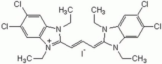
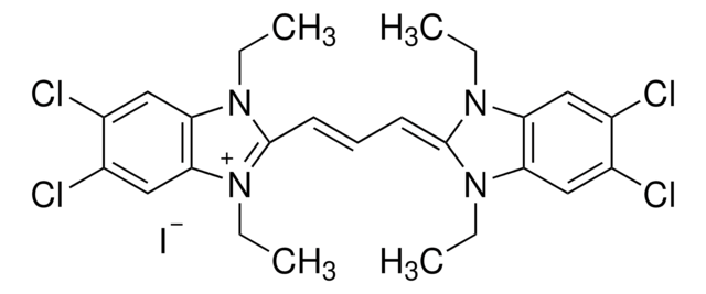
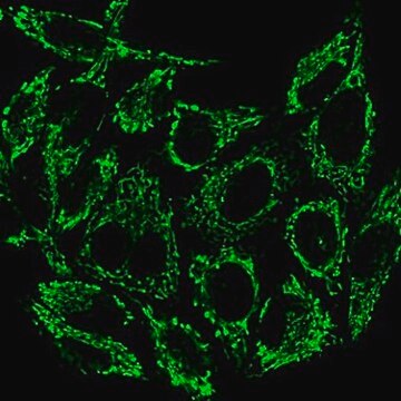
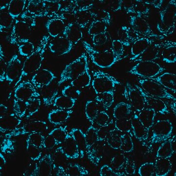
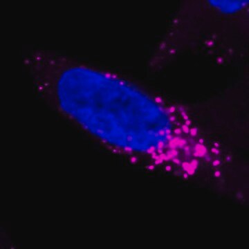
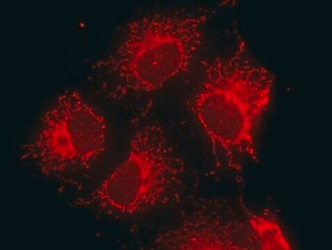
In normal cells, the JC-1 dye concentrates in the mitochondrial matrix where it forms red fluorescent aggregates. Any event that dissipates the mitochondrial membrane potential (e.g. apoptosis) prevents the accumulation of the JC-1 dye in the mitochondria and thus, the dye is dispersed throughout the entire cell leading to a shift from red (JC-1-aggregates) to green fluorescence (JC-1 monomers). The fluorescence of the cells stained with this kit may be observed by fluorescence microscopy or measured by fluorimetric and flow cytometry analysis.

Application

The dissipation of the mitochondrial electrochemical potential gradient (Δψ) is known as an early event in apoptosis. This kit offers a fast and convenient method for the detection of changes in mitochondrial inner-membrane electrochemical potential in living cells using the cationic, lipophilic dye, JC-1.

Features and Benefits

Kit offers:
  • A fast, simple, and convenient method for staining and assaying mitochondria intactness
  • Contains all the reagents required for the detection of changes mitochondrial inner-membrane electrochemical potential
  • Contains the antibiotic valinomycin, which can be used as a control agent that prevents JC-1 aggregation
  • The shift in fluorescence is clearly detectable in both green and red channels
  • Suitable for adherent cells as well as cells in suspension
  • Has been tested with Jurkat, U-937, HeLa, NIH3T3 cells
  • The fluorescence signal can be observed by fluorescence microscopy or measured by flow cytometry

Kit Components Only

Product No.
Description

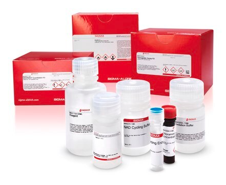
  • DMSO 1 mL

  • JC Staining Buffer 5X 120 mL

  • JC-1 1 mg

  • Valinomycin Ready Made .1 mL

Storage Class Code

10 - Combustible liquids

WGK

WGK 3

Flash Point(F)

Not applicable

Flash Point(C)

Not applicable


Certificates of Analysis (COA)

Search for Certificates of Analysis (COA) by entering the products Lot/Batch Number. Lot and Batch Numbers can be found on a product’s label following the words ‘Lot’ or ‘Batch’.

Already Own This Product?

Find documentation for the products that you have recently purchased in the Document Library.

Visit the Document Library

Customers Also Viewed

Slide 1 of 5

1 of 5

Small Molecule Mediated Restoration of Mitochondrial Function Augments Anti-Mycobacterial Activity of Human Macrophages Subjected to Cholesterol Induced Asymptomatic Dyslipidemia.
Suman Asalla et al.
Frontiers in cellular and infection microbiology, 7, 439-439 (2017-10-27)
Robert L Elliott et al.
PloS one, 14(4), e0214586-e0214586 (2019-04-02)
Many cells are cultured in media that contains an antibiotic to prevent bacterial contamination. Mycoplasma and other bacterial contamination is a serious problem for those involved in cell culture. Antibiotics in the media helps prevent this contamination and make life
Maria C Zanellati et al.
Frontiers in genetics, 6, 78-78 (2015-03-31)
Mutations in PARK2, encoding Parkin, cause an autosomal recessive form of juvenile Parkinson Disease (JPD). The aim of the present study was to investigate the impact of PARK2 mutations on mitochondrial function and morphology in human skin fibroblasts. We analyzed
Whasun Lim et al.
Journal of cellular physiology, 231(12), 2690-2699 (2016-03-13)
Apigenin is a flavonoid found in parsley, onions, oranges, tea, chamomile, wheat, and sprouts. It has a variety of biological properties including anti-oxidant, anti-mutagenic, anti-carcinogenic, anti-inflammatory, anti-proliferative, and anti-spasmodic effects. Based on epidemiological and case-control studies, apigenin is regarded as
A Cossarizza et al.
Biochemical and biophysical research communications, 197(1), 40-45 (1993-11-30)
A new method for the cytofluorimetric analysis of mitochondrial membrane potential in intact cells has been developed by using the lipophilic cationic probe 5,5',6,6'-tetrachloro-1,1',3,3'-tetraethylbenzimidazolcarbocyanine iodide (JC-1), whose monomer emits at 527 nm after excitation at 490 nm. Depending on the

Articles

Oxidative stress is mediated, in part, by reactive oxygen species produced by multiple cellular processes and controlled by cellular antioxidant mechanisms such as enzymatic scavengers or antioxidant modulators. Free radicals, such as reactive oxygen species, cause cellular damage via cellular.

Our team of scientists has experience in all areas of research including Life Science, Material Science, Chemical Synthesis, Chromatography, Analytical and many others.

Contact Technical Service