Skip to Content
Merck
All Photos(2)

Documents

C1926

Sigma-Aldrich

Monoclonal Anti-Collagen, Type IV antibody produced in mouse

clone COL-94, ascites fluid

Sign Into View Organizational & Contract Pricing


About This Item

MDL number:
UNSPSC Code:
12352203
NACRES:
NA.41

biological source

mouse

Quality Level

conjugate

unconjugated

antibody form

ascites fluid

antibody product type

primary antibodies

clone

COL-94, monoclonal

contains

15 mM sodium azide

species reactivity

human

technique(s)

dot blot: suitable
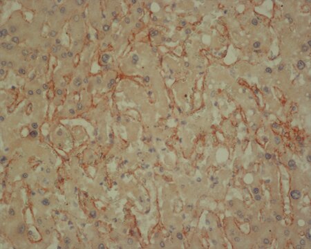
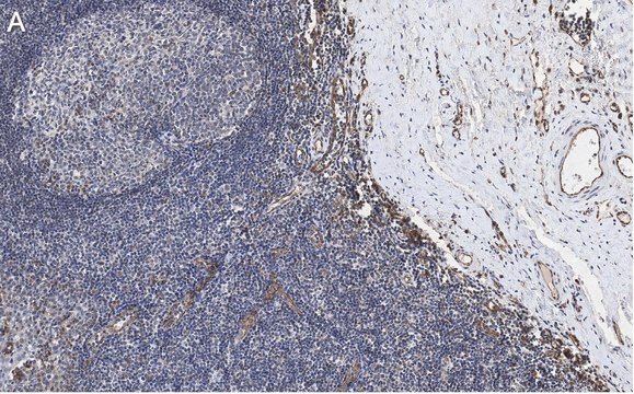
immunohistochemistry (formalin-fixed, paraffin-embedded sections): 1:500 using protease-digested sections of human tissue
immunohistochemistry (frozen sections): suitable
indirect ELISA: suitable
microarray: suitable

isotype

IgG1

UniProt accession no.

shipped in

dry ice

storage temp.

−20°C

target post-translational modification

unmodified

General description

Monoclonal Anti-Collagen Type IV (mouse IgG1 isotype) is derived from the COL-94 hybridoma produced by the fusion of mouse myeloma cells and splenocytes from BALB/c mice immunized with human collagen Type IV.
The composition of the extracellular matrix of all vertebrates is dominated by a class of molecules known as collagens, each with a unique feature suited to its function and location. Type IV collagen is a major constituent of basement membranes. This molecule is a continuous triple helix, about 400 nm in length, composed of at least two different α chain subunits termed α1 (IV) and α2 (IV) which may be composed of the same or different molecules. Upon proteolysis, fragments of 125 kDa, 95 kDa, and 60 kDa are released from a parent α1 (IV) chain, whereas fragments of 115 kDa, 80 kDa, and 45 kDa apparently originate from a distinct α2 (IV) chain.

Specificity

Using immunohistochemical staining of basal laminae, the antibody recognizes an epitope located on the α1 and/or α2 chains of human collagen type IV. The antibody shows reactivity with native primate collagen type IV in ELISA and dot blot assay. Using the dot blot technique, the antibody shows no cross-reactivity with human collagen types I, II, III, V, VI and VII, human vitronectin, fibronectin and chondroitin sulfate types A, B and C.

Immunogen

human collagen type IV

Application

Monoclonal Anti-Collagen, Type IV antibody produced in mouse has been used in
  • indirect immunolabeling
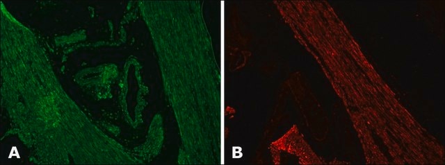
  • immunofluorescence staining
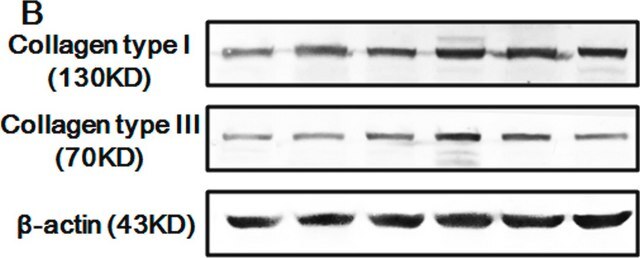
  • western blotting
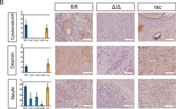
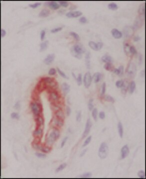
  • immunohistochemistry
  • immunocytochemistry

Biochem/physiol Actions

Collagens are known to have multiple functions. They are involved in tissue differentiation, morphogenesis and repair; play a role in the maintenance of tissue architecture; and form the filtration barrier of blood vessels.

Disclaimer

Unless otherwise stated in our catalog or other company documentation accompanying the product(s), our products are intended for research use only and are not to be used for any other purpose, which includes but is not limited to, unauthorized commercial uses, in vitro diagnostic uses, ex vivo or in vivo therapeutic uses or any type of consumption or application to humans or animals.

Not finding the right product?  

Try our Product Selector Tool.

Storage Class Code

12 - Non Combustible Liquids

WGK

nwg

Flash Point(F)

Not applicable

Flash Point(C)

Not applicable


Certificates of Analysis (COA)

Search for Certificates of Analysis (COA) by entering the products Lot/Batch Number. Lot and Batch Numbers can be found on a product’s label following the words ‘Lot’ or ‘Batch’.

Already Own This Product?

Find documentation for the products that you have recently purchased in the Document Library.

Visit the Document Library

The aromatase inhibitor anastrozole is associated with favorable embryo development and implantation markers in mice ovarian stimulation cycles
Karaer O, et al.
Fertility and Sterility, 83(6), 1797-1806 (2005)
Discoidin domain receptor 1 functions in axon extension of cerebellar granule neurons
Bhatt RS, et al.
Genes & Development, 14(17), 2216-2228 (2000)
Effect of single wall carbon nanotubes on human HEK293 cells
Cui D, et al.
Toxicology Letters, 155(1), 73-85 (2005)
Structure and Function of Basement Membranes
LeBleu VS et al
Exp. Biol. Med, 232, 1121-1129 (2007)
Aleksandra Kuzan et al.
Glycoconjugate journal, 35(1), 95-103 (2018-01-07)
The most abundant proteins in the arteries are those of extracellular matrix, ie. collagen and elastin. Due to their long half-lifes these proteins have an increased chance to undergo glycation. The aim of this study was to determine relationship between

Our team of scientists has experience in all areas of research including Life Science, Material Science, Chemical Synthesis, Chromatography, Analytical and many others.

Contact Technical Service