MABT395
Anti-Muc4 Antibody, clone 8G7
clone 8G7, from mouse
Synonym(s):
Mucin-4 beta chain
Sign Into View Organizational & Contract Pricing
All Photos(2)
About This Item
UNSPSC Code:
12352203
eCl@ss:
32160702
NACRES:
NA.41
Recommended Products
biological source
mouse
Quality Level
100
300
antibody form
purified immunoglobulin
antibody product type
primary antibodies
clone
8G7, monoclonal
species reactivity
human
technique(s)
immunohistochemistry: suitable
western blot: suitable
isotype
IgG1κ
UniProt accession no.
shipped in
wet ice
target post-translational modification
unmodified
Gene Information
human ... MUC4(4585)
Related Categories
General description
Mucin-4 (MUC-4 or Muc4) is also called Ascites sialoglycoprotein (ASGP), Pancreatic adenocarcinoma mucin, Testis mucin, and Tracheobronchial mucin. Muc4 is proteolytically cleaved into two chains: mucin-4 alpha chain and mucin-4 beta chain. Muc4 has anti-adhesive properties and is involved in tumor progression, cell proliferation and differentiation of epithelial cells. Muc4 is expressed in the thymus, thyroid, lung, trachea, esophagus, stomach, small intestine, colon, testis, prostate, ovary, uterus, placenta, mammary and salivary glands as well as lung cancers, squamous cell carcinomas of the upper aerodigestive tract, mammary carcinomas, biliary tract, colon, cervix cancers, pancreatic tumors, and pancreatic tumor cell lines. The expression of Muc4 has become a very useful indicator of poor prognosis in patients with invasive ductal carcinoma and intrahepatic cholangiocarcinoma, mass forming type (IDC,ICC-MF), where patients with high Muc4 expression had lower survival rates than those with low Muc4 expression.
Immunogen
KLH-conjugated linear peptide corresponding to the beta chain tandem repeat region of human Muc4.
Application
Anti-Muc4 Antibody, clone 8G7 is a highly specific mouse monoclonal antibody, that targets Mucin & has been tested in western blotting & IHC.
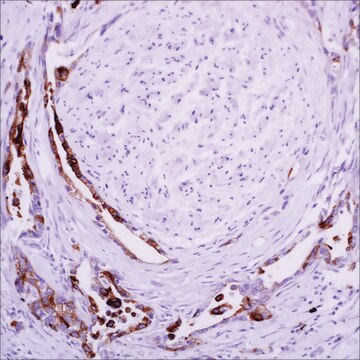
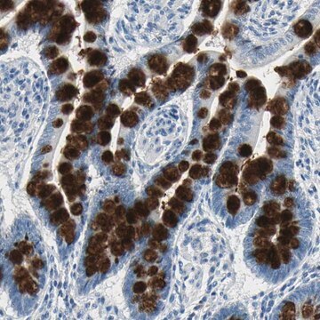
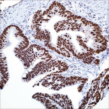
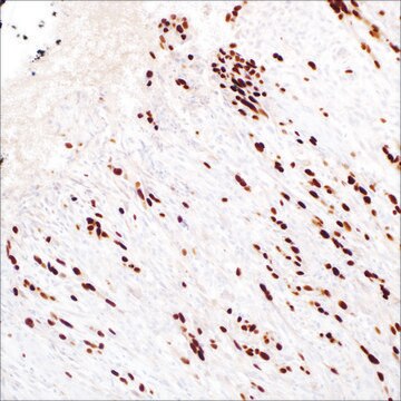
Immunohistochemistry Analysis: A 1:2,000 dilution from a representative lot detected Muc4 in human ilium tissues.
Western Blotting Analysis: A representative lot from an independent laboratory detected Muc4 in maligant melanoma and cutaneous squamous cell carcinoma tissue lysates (Moniaux, N., et al. (2004). J Histochem Cytochem. 52(2):253-261.).
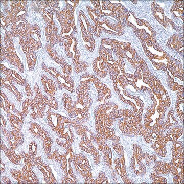
Immunohistochemistry Analysis: A representative lot from an independent laboratory detected Muc4 in lung adenocarcinoma and pancreatic adenocarcinoma tissues (Moniaux, N., et al. (2004). J Histochem Cytochem. 52(2):253-261.).
Immunohistochemistry Analysis: A representative lot from independent laboratories detected Muc4 in 330 tissue spots representing normal skin, and benign and malignant cutaneous diseases (Singh, A. P., et al. (2006). Prostate. 66(4):421-429.; Chakraborty, S., et al. (2010). J Clin Pathol. 63(7):579-584.).
Research Category
Cell Structure
Cell Structure
Research Sub Category
ECM Proteins
ECM Proteins
Quality
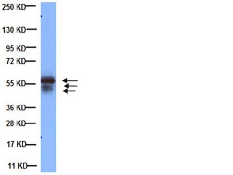
Evaluated by Western Blotting in CD18/HPAF cell lysate.
Western Blotting Analysis: 1 µg/mL of this antibody detected Muc4 in 10 µg of CD18/HPAF cell lysate.
Western Blotting Analysis: 1 µg/mL of this antibody detected Muc4 in 10 µg of CD18/HPAF cell lysate.
Target description
~240 kDa observed. Uniprot describes a molecular weight at 430 kDa; however, this protein may be observed at ~240 kDa due to alternative spicing and post-translational modification (Moniaux, N., et al. (2004). J Histochem Cytochem. 52(2):253-261.).
Physical form
Format: Purified
Protein G Purified
Purified mouse monoclonal IgG1κ in buffer containing 0.1 M Tris-Glycine (pH 7.4), 150 mM NaCl without preservatives.
Storage and Stability
Stable for 1 year at -20°C from date of receipt.
Handling Recommendations: Upon receipt and prior to removing the cap, centrifuge the vial and gently mix the solution. Aliquot into microcentrifuge tubes and store at -20°C. Avoid repeated freeze/thaw cycles, which may damage IgG and affect product performance.
Handling Recommendations: Upon receipt and prior to removing the cap, centrifuge the vial and gently mix the solution. Aliquot into microcentrifuge tubes and store at -20°C. Avoid repeated freeze/thaw cycles, which may damage IgG and affect product performance.
Other Notes
Concentration: Please refer to the Certificate of Analysis for the lot-specific concentration.
Disclaimer
Unless otherwise stated in our catalog or other company documentation accompanying the product(s), our products are intended for research use only and are not to be used for any other purpose, which includes but is not limited to, unauthorized commercial uses, in vitro diagnostic uses, ex vivo or in vivo therapeutic uses or any type of consumption or application to humans or animals.
Not finding the right product?
Try our Product Selector Tool.
WGK
WGK 1
Flash Point(F)
Not applicable
Flash Point(C)
Not applicable
Certificates of Analysis (COA)
Search for Certificates of Analysis (COA) by entering the products Lot/Batch Number. Lot and Batch Numbers can be found on a product’s label following the words ‘Lot’ or ‘Batch’.
Already Own This Product?
Find documentation for the products that you have recently purchased in the Document Library.
Our team of scientists has experience in all areas of research including Life Science, Material Science, Chemical Synthesis, Chromatography, Analytical and many others.
Contact Technical Service