General description
Tumor protein 63 (UniProt: Q9H3D4; also known as p63, Chronic ulcerative stomatitis protein, CUSP, Keratinocyte transcription factor KET, Transformation-related protein 63, TP63, Tumor protein p73-like, p73L, P40, P51) is encoded by the TP63 (also known as KET, P63, P73H, P73L, TP73L) gene (Gene ID: 8626) in human. TP63 is a member of the p53 protein family that is widely expressed. TP63 acts as a sequence specific DNA binding transcriptional activator or repressor. TP63 is shown to bind to DNA as a homotetramer and its DNA binding region is localized to amino acids 170-362. It has a transcription activation region (aa 1-107) and a transactivation inhibition region (aa 610-680), which can interact with, and inhibit the activity of the N-terminal transcriptional activation domain of TA-type isoforms. Twelve different isoforms of TP63 have been described that are produced by alternative promoter usage and alternative splicing. These isoforms contain a varying set of transactivation and auto-regulating transactivation inhibiting domains thus showing an isoform specific activity. The ratio of DeltaN-type and TA-type isoforms governs the maintenance of epithelial stem cell compartments and regulates the initiation of epithelial stratification from the undifferentiated embryonal ectoderm. Isoform 2 is reported to activate RIPK4 transcription and may be required in conjunction with TP73/p73 for initiation of p53/TP53 dependent apoptosis in response to genotoxic insults and the presence of activated oncogenes. Isoform 10 is predominantly expressed in skin squamous cell carcinoma, but not in normal skin tissue.
Specificity
Clone 11H1 detects human Tumor protein 63 (P40). It targets an epitope with in the first 205 amino acids from the N-terminal region.
Immunogen
Epitope: N-terminus
MBP-conjugated recombinant fragment corresponding to the first 205 amino acids from human Tumor protein 63.
Application
Anti-P40 (TP63), clone 11H1, Cat. No. MABT1497, is a mouse monoclonal antibody that detects Tumor protein 63 and has been tested for use in Western Blotting.
Research Category
Epigenetics & Nuclear Function
Quality
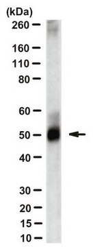
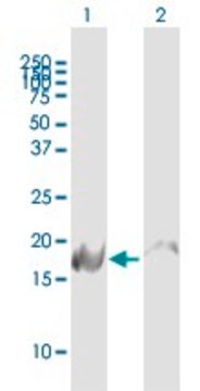
Evaluated by Western Blotting in A431 cell lysate.
Western Blotting Analysis: A 1:250 dilution of this antibody detected P40 (TP63) in A431 cell lysate.
Target description
~66 kDa observed; 76.78 kDa calculated.Uncharacterized bands may be observed in some lysate(s).
Physical form
Format: Purified
Protein G purified
Purified mouse monoclonal antibody IgG1 in buffer containing 0.1 M Tris-Glycine (pH 7.4), 150 mM NaCl with 0.05% sodium azide.
Storage and Stability
Stable for 1 year at 2-8°C from date of receipt.
Other Notes
Concentration: Please refer to lot specific datasheet.
Disclaimer
Unless otherwise stated in our catalog or other company documentation accompanying the product(s), our products are intended for research use only and are not to be used for any other purpose, which includes but is not limited to, unauthorized commercial uses, in vitro diagnostic uses, ex vivo or in vivo therapeutic uses or any type of consumption or application to humans or animals.

