Skip to Content
Merck
All Photos(1)

Documents

07-416

Sigma-Aldrich

Anti-Akt1/PKBα Antibody

Upstate®, from rabbit

Synonym(s):

Protein kinase B, RAC-alpha serine/threonine-protein kinase, murine thymoma viral (v-akt) oncogene homolog-1, rac protein kinase alpha, v-akt murine thymoma viral oncogene homolog 1

Sign Into View Organizational & Contract Pricing


About This Item

UNSPSC Code:
12352203
eCl@ss:
32160702
NACRES:
NA.41

biological source

rabbit

Quality Level

antibody form

purified antibody

antibody product type

primary antibodies

clone

polyclonal

species reactivity

rat, human, mouse

packaging

antibody small pack of 25 μg

manufacturer/tradename

Upstate®

technique(s)

immunoprecipitation (IP): suitable
western blot: suitable

isotype

IgG

NCBI accession no.

UniProt accession no.

shipped in

ambient

target post-translational modification

unmodified

Gene Information

human ... AKT1(207)

Related Categories

General description

Akt is the major known effector of the PI3 Kinase pathway. Generation of PIP3 results in the activation of PDK1, which phosphorylates Akt on Thr308, and another kinase complex, thought to be the mTORC2 complex (or possibly an intermediate kinase linking the two) that phosphorylates Akt on Ser473. These phosphorylations additively activate Akt Ser/Thr kinase activity, and the use of phosphorylation state-specific antibodies directed against either of these sites can imply Akt activation. Activation of Akt can be measured directly by immunoprecipitation followed by phosphorylation of a known substrate with radiolabeled ATP. Akt has been shown to phosphorylate over 70 substrates including TSC1&2 in the mTOR pathway, Bad on Ser136, GSK3, which is inactivated by this phosphorylation, the FoxO family of transcription factors, PRAS40, AS160.

Specificity

Recognizes Akt1/PKBα, MW ~60 kDa.

Immunogen

15 residue synthetic peptide (C-RPHFPQFS YSASGTA) corresponding to the C-terminal (residues 466-480) of rat Akt1, with an N-terminal cysteine added for conjugation purposes. The Akt1 sequence is identical to mouse Akt1 (15/15 amino acids) and strongly conserved in bovine Akt1 (14/15 amino acids) and human Akt1 (13/15 amino acids). Human Akt2 shares 11/15 amino acids with the peptide immunogen sequence.
Epitope: C-terminus

Application

Immunoprecipitation Kinase Assay:
4 µg of a previous lot immunoprecipitated active Akt from 1 mg of HEK293 cells stimulated with IGF-1.
Research Category
Signaling
Research Sub Category
PI3K, Akt, & mTOR Signaling
This Anti-Akt1/PKBα Antibody is validated for use in IP, WB for the detection of Akt1/PKBα.

Quality

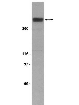
Evaluated by western blot in RIPA lysates from human A431 carcinoma cells.

Western Blot Analysis:
0.5-2 µg/mL of this antibody detected Akt in RIPA lysates from human A431 carcinoma cells.

Target description

~60 kDa

Physical form

Format: Purified
Protein A purified
Purified rabbit polyclonal in buffer containing 0.1 M Tris-glycine, pH 7.4, 0.15 M NaCl, 0.05% sodium azide before the addition of glycerol to 30%.

Storage and Stability

Stable for 1 year at -20ºC from date of receipt.

Analysis Note

Control
Positive Antigen Control: Catalog #12-301, non-stimulated A431 cell lysate. Add 2.5µL of 2-mercaptoethanol/100µL of lysate and boil for 5 minutes to reduce the preparation. Load 20µg of reduced lysate per lane for minigels.

Other Notes

Concentration: Please refer to the Certificate of Analysis for the lot-specific concentration.

Legal Information

UPSTATE is a registered trademark of Merck KGaA, Darmstadt, Germany

Disclaimer

Unless otherwise stated in our catalog or other company documentation accompanying the product(s), our products are intended for research use only and are not to be used for any other purpose, which includes but is not limited to, unauthorized commercial uses, in vitro diagnostic uses, ex vivo or in vivo therapeutic uses or any type of consumption or application to humans or animals.

Not finding the right product?  

Try our Product Selector Tool.

WGK

WGK 1


Certificates of Analysis (COA)

Search for Certificates of Analysis (COA) by entering the products Lot/Batch Number. Lot and Batch Numbers can be found on a product’s label following the words ‘Lot’ or ‘Batch’.

Already Own This Product?

Find documentation for the products that you have recently purchased in the Document Library.

Visit the Document Library

Angela M Whetzel et al.
Circulation research, 99(7), 731-739 (2006-09-09)
Monocyte recruitment and adhesion to vascular endothelium are key early events in atherosclerosis. We examined the role of sphingosine-1-phosphate (S1P) on modulating monocyte/endothelial interactions in the NOD/LtJ (NOD) mouse model of type 1 diabetes. Aortas from nondiabetic and diabetic NOD
Francesco Papaleo et al.
The Journal of neuroscience : the official journal of the Society for Neuroscience, 36(17), 4859-4875 (2016-04-29)
Schizophrenia is a chronic, disabling neuropsychiatric disorder with complex genetic origins. The development of strategies for genome manipulation in rodents provides a platform for understanding the pathogenic role of genes and for testing novel therapeutic agents. Neuregulin 1 (NRG1), a
Dose-dependent effects of sirolimus on mTOR signaling and polycystic kidney disease.
Novalic, Z; van der Wal, AM; Leonhard, WN; Koehl, G; Breuning, MH; Geissler, EK; de Heer et al.
Journal of the American Society of Nephrology null
Heidi Hynynen et al.
Physiological reports, 8(13), e14474-e14474 (2020-07-10)
Dietary fats are essential for cardiac function. The metabolites of fats known as fatty acids provide most of the energy for cardiac tissue, serve as building blocks for membranes and regulate important signaling cascades. Despite their importance, excess fat intake
Y Sei et al.
Molecular psychiatry, 12(10), 946-957 (2007-04-19)
Neuregulin1 (NRG1), a candidate susceptibility gene for schizophrenia, plays a critical role in neuronal migration and central nervous system development. However, its relation to schizophrenia pathogenesis is unknown. Here we show that B lymphoblasts migrate to NRG1 through the ErbB-signaling

Articles

Autophagy is a highly regulated process that is involved in cell growth, development, and death. In autophagy cells destroy their own cytoplasmic components in a very systematic manner and recycle them.

Our team of scientists has experience in all areas of research including Life Science, Material Science, Chemical Synthesis, Chromatography, Analytical and many others.

Contact Technical Service